Monday, to King's, to have cardiac ambulatory test. Three electrodes are strapped to the chest, and the signals are collected on a rectangular device about the size of a cigarette packet, over a period of 24 hours.
MRI of the brain didn't show any acute ischaemic lesions but there is a background of moderate small vessel disease. There are some discrete white matter lesions which appear to be more numerous on the left than on the right. Carotid Dopplers showed 40-49% on the right and 20-30% on the left. Interestingly, the white matter lesions on the brain are more numerous on the side of the lower grade carotid stenosis.
Regarding the EEG, the background activity shows a bilateral and symmetrical alpha, at 8-9 Hz, up to 78 μV.It is seen over posterior regions and attenuates on visual attention. Some slow theta and delta activity is seen over temporal regions, especially over the left hemisphere. Some sharp transients may be seen associated to slow waves, and seen over fronto-temporal regions as well as more prominent over the left hemisphere. These probably don't represent epileptiform discharges but rather, non-specific changes which are seen in cerebro-vascular disease.
Francis Story The Search for the Self
The search for Self is vain, because it is a search for something that does not exist except as a mythical concept which has had to be taken into the structure of language by common assent. If it is used in any other way than as a fictitious convenience - if it is taken as meaning something real and enduring - it cannot be anything but a stumbling-block to the development of right understanding.
Blog Archive
-
▼
2010
(439)
-
▼
December
(27)
- Hate crime against Ahmadis
- Friday
- Wednesday
- My teenage visitors
- Short week
- No title
- No title
- Karen Dobrowska
- No title
- No title
- No title
- Tuesday morning, I chaired a seminar on the catas...
- Last Saturday my cousin Dudi died at the age of 92...
- Haemoglobin readings from blood tests
- Tuesday, meeting of Parliamentary Human Rights Gro...
- Haemoglobin readins from blood tests
- Monday, to King's, to have cardiac ambulatory test...
- Rest of the week
- Tuesday continued
- Introducing the Peru Roundtable
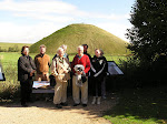
- From left to right: Jose de Echave, Anne Lindsay,...
- Going back to Tuesday, the morning was taken up ch...
- The garden, from eye level
- Flodden Road, from our bedroom window this mornin...
- December 2, from JW's window
-
▼
December
(27)
About Me
- Eric Avebury
- Eric Lubbock, Lord Avebury, b September 29, 1928. Upper Canada College & Balliol College Oxford (BA 1949, boxing blue); Welsh Guards (Second Lieut) 1949-51; Rolls Royce (aero-engine division) 1951-6; Production Engineering 1956-60; Charterhouse Group 1960-2. MP Orpington 1962-70; Liberal Chief Whip 1963-70; Chair, Parliamentary Civil Liberties Group 1964-70; Parliamentary Human Rights Group, 1976-1997; Traveller Law Reform Unit; Peru Support Group, 2003-; Cameroon Campaign Group 2003- Speaker's Conference on Electoral Law 1963-5; Select Committee on Science and Technology, 1968-70; Royal Commission on Standards of Conduct in Public Life, 1974-6 President, Data Processing Management Association, 1972-5; Fluoridation Society, 1972-84; Conservation Society, 1973-83; London Bach Society, 1984-98; ACERT (Advisory Council for Education of Romanies & Travellers) 2001-;TAPOL (Indonesian human rights); Kurdish Human Rights Project; Patron, Angulimala (Buddhist Prison Chaplaincy), 1992-; Founder, Parliamentarians for East Timor, 1988; Vice-Chair, Parliamentary Group for Tibet; Member, Institution of Mechanical Engineers (MIMechE); Fellow, British Computer Society (FBCS).
No comments:
Post a Comment